


Exophthalmos of the eye in an 11 year old cat. Diagnosis and conclusions
An 11-year-old European cat is presented for consultation due to the appearance of a conjunctival mass, exophthalmos, and various corneal lesions in the left eye. The exophthalmos has notably led to lagophthalmos, resulting in corneal ulcer and inflammation. However, the general condition of the animal remains good.
Exophthalmos at the clinic
Following the clinical examination, a diagnosis has not yet been established. A corneal ulcer is identified using fluorescein, but it is secondary to lagophthalmos. A request for an ophthalmology consultation on the app DRVET.CH is sent with a photo taken during the examination and the important clinical findings mentioned above

Veterinarian sends a consultation request to an ophthalmologist through the DRVET.CH app with a photo and examination results
Remote ophthalmological consultation
After the consultation, several tests are recommended. Conjunctival cytology is performed using a cytobrush and local anaesthesia. An ultrasound is also recommended, which does not show any intraocular abnormalities, and the retrobulbar area is difficult to evaluate. The cytology is sent to the laboratory and returns inconclusive. As exophthalmos is progressing rapidly, a decision to perform a cranial scan to explore the cause has been made with the owners.
At the end of the consultation, a conservative treatment with antibiotic ointment (Tobrex, tobramycin) and ocular lubricant (Remend cornea, hyaluronic acid) is initiated.
At the end of the consultation, a conservative treatment with antibiotic ointment (Tobrex, tobramycin) and ocular lubricant (Remend cornea, hyaluronic acid) is initiated.
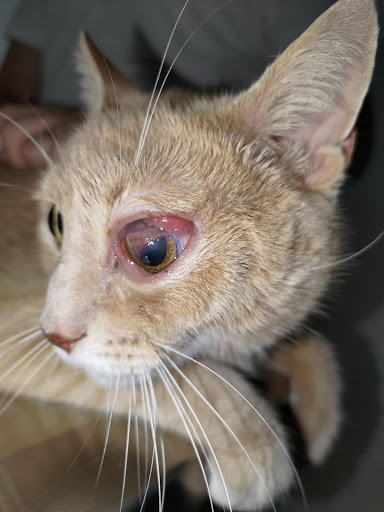
An 11-year-old cat with conjunctivitis, exophthalmos and various corneal lesions in the left eye
Results of the scan
The scan reveals a large tissue mass extending into the orbital tissues around the periphery of the left eye, encircling it, with an appearance consistent with an aggressive primary tumor. This mass also infiltrates the left trigeminal nerve. Fine needle aspirates are performed and unfortunately suggest a high-grade lymphom
Fig1. Hyperintense tissue swelling located at the periphery of the left eyeball on T2 FLAIR weighting, causing a mass effect on the left eyeball.
Fig 2. Thickening of the left trigeminal nerve with pronounced enhancement.


Lymphoma diagnosis
Ocular lymphomas in cats are tumors that can take on various forms and affect different structures of the eye. They are the second most common ocular tumor in the feline species. The lack of characteristic presentations can make their diagnosis more challenging. Unfortunately, the prognosis is poor, but chemotherapy and adjunctive treatments can help maintain a good quality of life for the animal.


Ready to communicate?
Yes, I want to join DRVET
Pierre Starkov
Co-founder DRVET

Daniel Pereira
Co-founder DRVET